
To understand the anatomy of the ear, we follow sound as it gets picked up by the outer ear, and is transmitted through the middle ear to the inner ear. 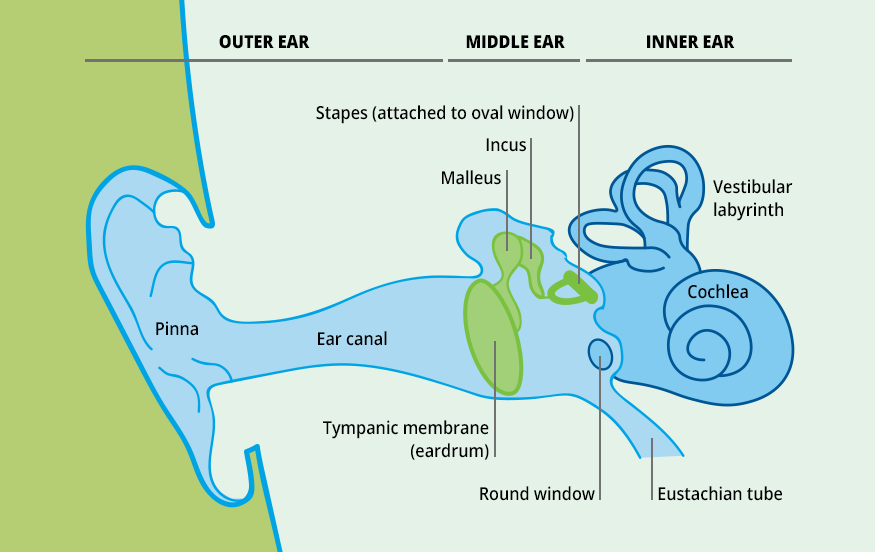
Outer Ear Anatomy
The outer ear consists of the pinna and the external auditory canal, also called the ear canal.
LEARN MORE:
LISTENING IN 3D
As already discussed in the article ‘Listening in 3D’, the pinna plays an important role in the external auditory source location. Also, its horn-like shape provides a smooth transition from the ‘infinite’ space around the head, funneling the sound into the narrow auditory canal.
The external acoustic meatus (also called external auditory meatus) then guides the sound towards the eardrum, a thin membrane separating the outer from the middle ear.
Middle Ear Anatomy
The middle ear is a small air-filled chamber between the outer and inner ear. The purpose of this chamber is twofold. First, it contains a mechanism of three tiny bones / small bones, called the auditory ossicles, connecting the eardrum and the inner ear. This gearbox like mechanism is needed since the inner ear is filled with a fluid, making direct excitation by the eardrum inefficient.
Secondly, the middle ear is needed to equalize pressure across the eardrum, also called the tympanic membrane. A healthy eardrum is completely airtight, preventing airflow from the outer ear into the middle ear. The pressure difference between the two chambers moves the membrane in and out, which is exactly what is needed to pick up the rapid pressure fluctuations of sound waves.
Outer ear dimensions and amplification
Acoustically, the outer ear works as a tube resonator, with the strongest first resonance around 3 kHz, where a quarter wavelength of sound in air (10 cm / 4 = 2.5 cm) fits the length of the ear canal. In contrast, sensitivity drops significantly at lower frequencies where the wavelengths are large compared to the ear’s size.
MICROPHONES
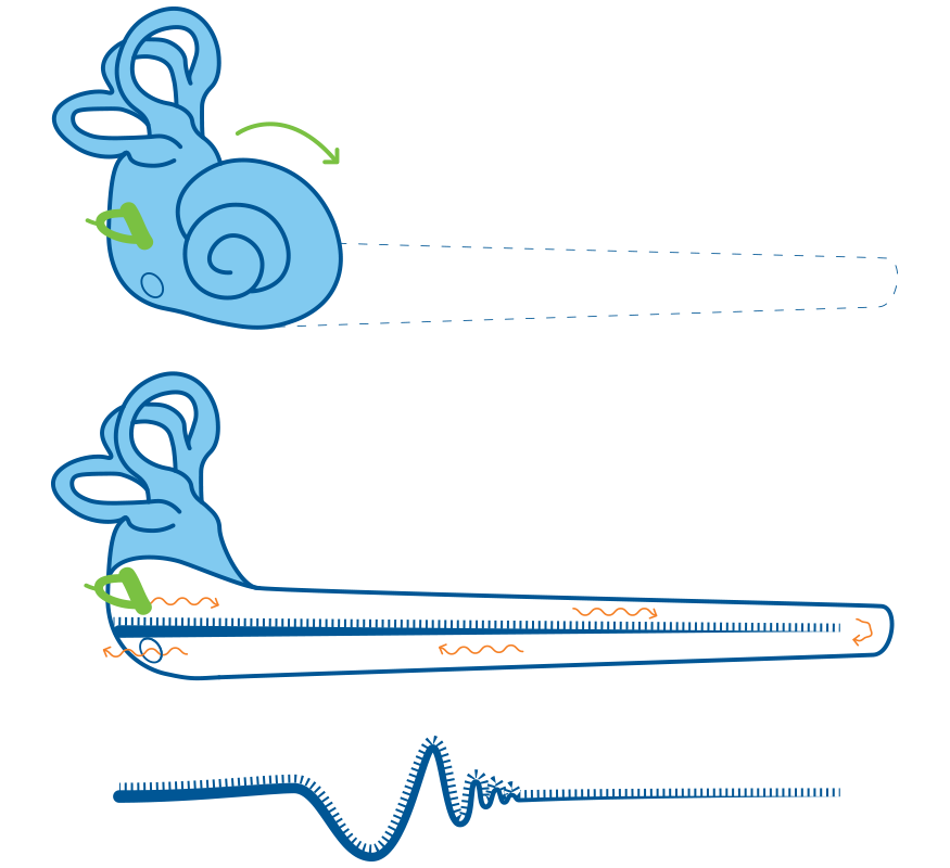
Cochlea with the basilar membrane
Even when excited by the sound of a pure tone, the entire basilar membrane will be set into motion. However, the area associated with the frequency will react the most; that is, the lateral oscillations will peak around this section.
However, a problem can arise when the atmospheric (static) pressure in the outer ear differs from the pressure inside the middle ear.
This mechanism is not that evident in everyday life but is easily experienced during lift-off and landing on an airplane, where the ambient pressure changes significantly due to the change in altitude. The pressure in the outer ear follows the ambient pressure in the airplane, whereas the pressure on the inside of the eardrum remains unchanged. The constant pressure difference applies a pre-tension to the membrane, pushing it either in or out, which gives an unpleasant sensation and leads to sound being perceived duller.
The Eustachian tube, which connects the middle ear to the throat, helps to equalize the sound pressure.
When we swallow, the tube opens briefly causing the static pressure on the inside of the eardrum to equalize to that of the outer ear, resetting the eardrum to its neutral position. The eardrum will have its normal sensitivity and sound will again be bright.
Inner Ear Anatomy
The inner ear is the most complex element in the chain. It is a fluid-filled chamber and consists of two parts: the vestibular labyrinth, which functions as part of the body’s balance mechanism, and the cochlea, containing the basilar membrane and the hearing organ of Corti, a sensory element that converts and enables the transfer of sound waves into nerve impulses so that our brain can process the information.
Sound Pressure Equalization In A Microphone Diaphragm
To convert sound pressure into an electrical signal, Brüel & Kjær’s condenser microphones use a delicate diaphragm stretched across a backplate with a very narrow gap between them, forming a capacitor.
Impinging sound deflects the diaphragm, and the variation in distance to the backplate produces an electrical signal proportional to the sound pressure. The diaphragm seals the microphone at the top so that a variation in the static, ambient pressure would change the diaphragm’s neutral position relative to the backplate.
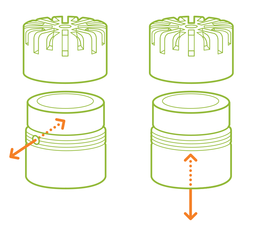
The ear solves this problem with the Eustachian tube, and condenser microphones use a similar design. A narrow air channel at the side or rear of the microphone ensures that the internal cavity’s static pressure equalizes with the environment.
This hearing organ contains thousands of small hair cells, which are connected to the acoustic nerve. The oscillation pattern of the basilar membrane is quite complex, with different areas being stimulated more or less by different frequencies. For each of these areas, a different group of hair cells will be activated and send impulses through the nerves to the brain. Thus, the organ of Corti splits up the sound into its spectral components, similar to raindrops splitting up sunlight into individual colors.
Matthias Scholz
User Interface Designer
Ph.D. Applied Acoustics
Hottinger Brüel & Kjær
Well, at least this is the short version.
The long version is much more complex, but also exciting, explaining the reason behind many of the phenomena, we experience in our perception of sound.
It deserves a separate chapter, which you can find here: Anatomy of the Human Ear - Part 2
뉴스레터를 구독하고 소리와 진동에 대한 최신 이야기를 만나보세요
Brüel & Kjær가 전하는 최신 뉴스가 받은편지함으로 발송되었습니다.
신제품 출시, 할인 및 이벤트
소음 진동 기사, 비디오 및 가이드